How Does A Plant Cell Look Like Under The Microscope . Zooming in even closer to the cell wall structure, the slcu microscopy facility collaborated with plant scientists in paris, discovering new filamentous structures within plant cell walls that help plants build their complex shapes. In all cases, the image shows a seed coat cell of a developing seed of the model plant, arabidopsis. The central vacuole is a large organelle that often fills most of the plant cell. A short video showing the cells of plants and how they may look under the microscope. Image in (a) is a light microscopy image,. The most common specimens to observe under a light microscope are cheek cells (animal cells) and onion cells (plant cells) a stain is often used to ensure cell structures are clearly visible. Looking at the structure of cells in the microscope. It is filled with liquid and surrounded by a.
from www.vecteezy.com
Zooming in even closer to the cell wall structure, the slcu microscopy facility collaborated with plant scientists in paris, discovering new filamentous structures within plant cell walls that help plants build their complex shapes. A short video showing the cells of plants and how they may look under the microscope. The most common specimens to observe under a light microscope are cheek cells (animal cells) and onion cells (plant cells) a stain is often used to ensure cell structures are clearly visible. Looking at the structure of cells in the microscope. Image in (a) is a light microscopy image,. It is filled with liquid and surrounded by a. The central vacuole is a large organelle that often fills most of the plant cell. In all cases, the image shows a seed coat cell of a developing seed of the model plant, arabidopsis.
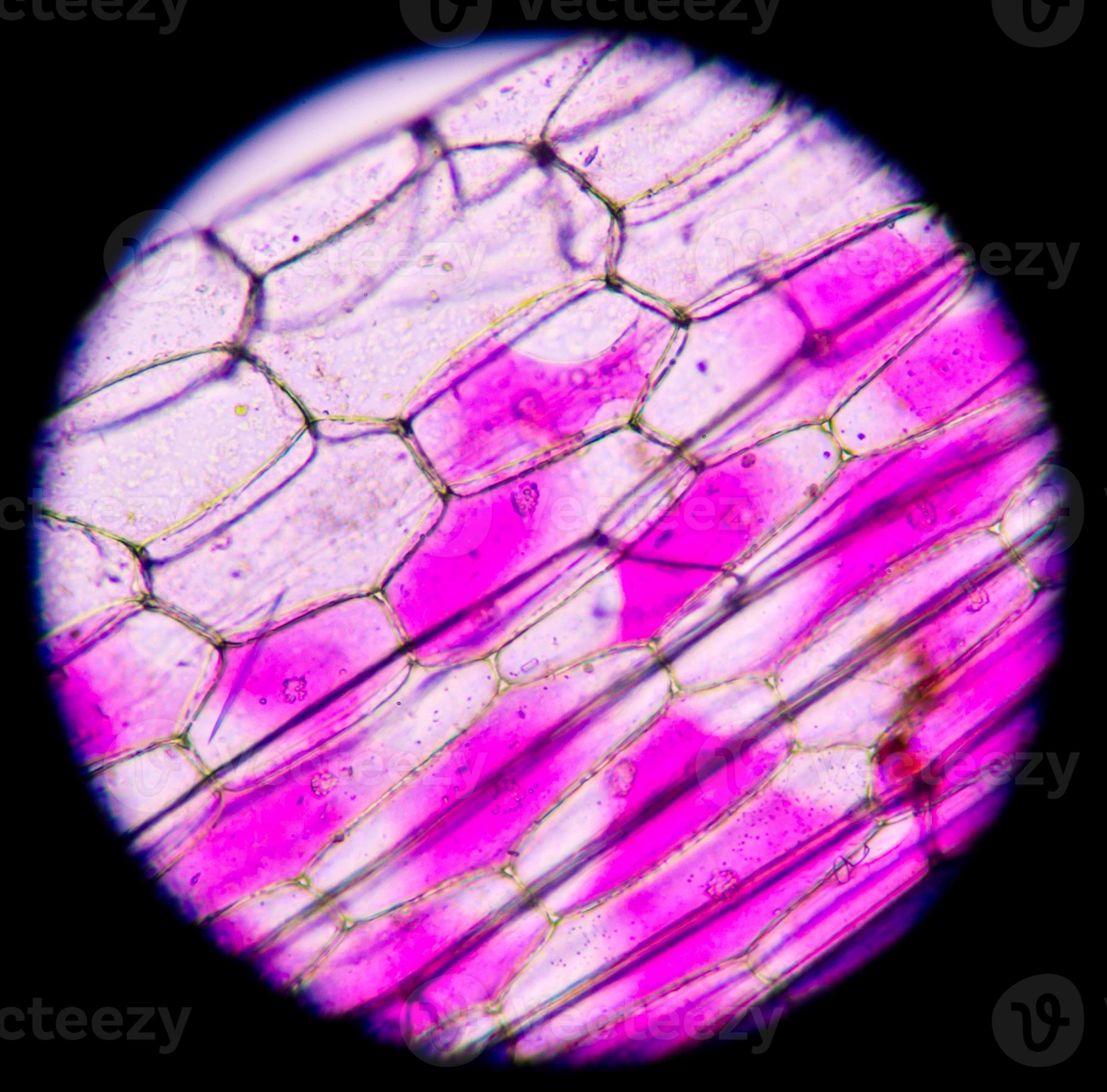
plant cells under microscope.400x 937256 Stock Photo at Vecteezy
How Does A Plant Cell Look Like Under The Microscope The central vacuole is a large organelle that often fills most of the plant cell. Zooming in even closer to the cell wall structure, the slcu microscopy facility collaborated with plant scientists in paris, discovering new filamentous structures within plant cell walls that help plants build their complex shapes. In all cases, the image shows a seed coat cell of a developing seed of the model plant, arabidopsis. Looking at the structure of cells in the microscope. It is filled with liquid and surrounded by a. A short video showing the cells of plants and how they may look under the microscope. The central vacuole is a large organelle that often fills most of the plant cell. Image in (a) is a light microscopy image,. The most common specimens to observe under a light microscope are cheek cells (animal cells) and onion cells (plant cells) a stain is often used to ensure cell structures are clearly visible.
From room201csa6grade.weebly.com
6 TH GRADERS Science How Does A Plant Cell Look Like Under The Microscope The central vacuole is a large organelle that often fills most of the plant cell. Zooming in even closer to the cell wall structure, the slcu microscopy facility collaborated with plant scientists in paris, discovering new filamentous structures within plant cell walls that help plants build their complex shapes. Image in (a) is a light microscopy image,. The most common. How Does A Plant Cell Look Like Under The Microscope.
From www.pinterest.nz
Epidermal onion cells under a microscope. Plant cells appear polygonal How Does A Plant Cell Look Like Under The Microscope Image in (a) is a light microscopy image,. Zooming in even closer to the cell wall structure, the slcu microscopy facility collaborated with plant scientists in paris, discovering new filamentous structures within plant cell walls that help plants build their complex shapes. The central vacuole is a large organelle that often fills most of the plant cell. A short video. How Does A Plant Cell Look Like Under The Microscope.
From blog.mozilla.com.tw
Plant Cell Printable How Does A Plant Cell Look Like Under The Microscope It is filled with liquid and surrounded by a. The most common specimens to observe under a light microscope are cheek cells (animal cells) and onion cells (plant cells) a stain is often used to ensure cell structures are clearly visible. The central vacuole is a large organelle that often fills most of the plant cell. Looking at the structure. How Does A Plant Cell Look Like Under The Microscope.
From franklynnovencidoe02737.blogspot.com
Plant Cell Under Microscope Labeled Pin By Nia On Education Plant How Does A Plant Cell Look Like Under The Microscope In all cases, the image shows a seed coat cell of a developing seed of the model plant, arabidopsis. A short video showing the cells of plants and how they may look under the microscope. The most common specimens to observe under a light microscope are cheek cells (animal cells) and onion cells (plant cells) a stain is often used. How Does A Plant Cell Look Like Under The Microscope.
From mavink.com
Plant Cell Light Microscope How Does A Plant Cell Look Like Under The Microscope The most common specimens to observe under a light microscope are cheek cells (animal cells) and onion cells (plant cells) a stain is often used to ensure cell structures are clearly visible. Zooming in even closer to the cell wall structure, the slcu microscopy facility collaborated with plant scientists in paris, discovering new filamentous structures within plant cell walls that. How Does A Plant Cell Look Like Under The Microscope.
From www.reddit.com
Plant cells under the microscope r/MicroPorn How Does A Plant Cell Look Like Under The Microscope In all cases, the image shows a seed coat cell of a developing seed of the model plant, arabidopsis. The most common specimens to observe under a light microscope are cheek cells (animal cells) and onion cells (plant cells) a stain is often used to ensure cell structures are clearly visible. The central vacuole is a large organelle that often. How Does A Plant Cell Look Like Under The Microscope.
From www.animalia-life.club
Eukaryotic Cells Microscope How Does A Plant Cell Look Like Under The Microscope It is filled with liquid and surrounded by a. The central vacuole is a large organelle that often fills most of the plant cell. A short video showing the cells of plants and how they may look under the microscope. Zooming in even closer to the cell wall structure, the slcu microscopy facility collaborated with plant scientists in paris, discovering. How Does A Plant Cell Look Like Under The Microscope.
From clairciccarelloe03349.blogspot.com
Plant Cell Under Electron Microscope Labelled / Animal Cells and Plant How Does A Plant Cell Look Like Under The Microscope In all cases, the image shows a seed coat cell of a developing seed of the model plant, arabidopsis. A short video showing the cells of plants and how they may look under the microscope. Image in (a) is a light microscopy image,. The central vacuole is a large organelle that often fills most of the plant cell. It is. How Does A Plant Cell Look Like Under The Microscope.
From www.youtube.com
Plant Cells Under Compound Microscope Biology Practical YouTube How Does A Plant Cell Look Like Under The Microscope The central vacuole is a large organelle that often fills most of the plant cell. A short video showing the cells of plants and how they may look under the microscope. Looking at the structure of cells in the microscope. The most common specimens to observe under a light microscope are cheek cells (animal cells) and onion cells (plant cells). How Does A Plant Cell Look Like Under The Microscope.
From www.dreamstime.com
Plant Cell Under the Microscope View Stock Photo Image of botany How Does A Plant Cell Look Like Under The Microscope A short video showing the cells of plants and how they may look under the microscope. The most common specimens to observe under a light microscope are cheek cells (animal cells) and onion cells (plant cells) a stain is often used to ensure cell structures are clearly visible. In all cases, the image shows a seed coat cell of a. How Does A Plant Cell Look Like Under The Microscope.
From twitter.com
Krystal Clover on Twitter "How do plant cells look under a microscope How Does A Plant Cell Look Like Under The Microscope Image in (a) is a light microscopy image,. In all cases, the image shows a seed coat cell of a developing seed of the model plant, arabidopsis. The central vacuole is a large organelle that often fills most of the plant cell. Looking at the structure of cells in the microscope. It is filled with liquid and surrounded by a.. How Does A Plant Cell Look Like Under The Microscope.
From www.vrogue.co
Fajarv Prophase Plant Cell Under Microscope vrogue.co How Does A Plant Cell Look Like Under The Microscope The most common specimens to observe under a light microscope are cheek cells (animal cells) and onion cells (plant cells) a stain is often used to ensure cell structures are clearly visible. The central vacuole is a large organelle that often fills most of the plant cell. A short video showing the cells of plants and how they may look. How Does A Plant Cell Look Like Under The Microscope.
From nghenhansu.edu.vn
Collection 92+ Images What Does A Plant Cell Look Like Under A How Does A Plant Cell Look Like Under The Microscope Looking at the structure of cells in the microscope. The central vacuole is a large organelle that often fills most of the plant cell. In all cases, the image shows a seed coat cell of a developing seed of the model plant, arabidopsis. It is filled with liquid and surrounded by a. Image in (a) is a light microscopy image,.. How Does A Plant Cell Look Like Under The Microscope.
From plantideas.darienicerink.com
What Does A Plant Cell Look Like Plant Ideas How Does A Plant Cell Look Like Under The Microscope Image in (a) is a light microscopy image,. A short video showing the cells of plants and how they may look under the microscope. Zooming in even closer to the cell wall structure, the slcu microscopy facility collaborated with plant scientists in paris, discovering new filamentous structures within plant cell walls that help plants build their complex shapes. It is. How Does A Plant Cell Look Like Under The Microscope.
From animalia-life.club
Plant Cell Nucleus Microscope How Does A Plant Cell Look Like Under The Microscope A short video showing the cells of plants and how they may look under the microscope. Looking at the structure of cells in the microscope. In all cases, the image shows a seed coat cell of a developing seed of the model plant, arabidopsis. It is filled with liquid and surrounded by a. Image in (a) is a light microscopy. How Does A Plant Cell Look Like Under The Microscope.
From clairciccarelloe03349.blogspot.com
Plant Cell Under Electron Microscope Labelled / Animal Cells and Plant How Does A Plant Cell Look Like Under The Microscope The most common specimens to observe under a light microscope are cheek cells (animal cells) and onion cells (plant cells) a stain is often used to ensure cell structures are clearly visible. Looking at the structure of cells in the microscope. Image in (a) is a light microscopy image,. In all cases, the image shows a seed coat cell of. How Does A Plant Cell Look Like Under The Microscope.
From mungfali.com
Plant Cell Under Compound Microscope How Does A Plant Cell Look Like Under The Microscope The most common specimens to observe under a light microscope are cheek cells (animal cells) and onion cells (plant cells) a stain is often used to ensure cell structures are clearly visible. Image in (a) is a light microscopy image,. The central vacuole is a large organelle that often fills most of the plant cell. It is filled with liquid. How Does A Plant Cell Look Like Under The Microscope.
From www.pinterest.jp
PlantStemSection Microscopic photography, Things under a microscope How Does A Plant Cell Look Like Under The Microscope In all cases, the image shows a seed coat cell of a developing seed of the model plant, arabidopsis. Zooming in even closer to the cell wall structure, the slcu microscopy facility collaborated with plant scientists in paris, discovering new filamentous structures within plant cell walls that help plants build their complex shapes. It is filled with liquid and surrounded. How Does A Plant Cell Look Like Under The Microscope.